
Author: Stephanie Thomas, CVT, VTS (Anesthesia Clinical Manager)
Monitoring blood pressure during anesthesia is paramount to ensuring patient safety. At PASE, we often employ advanced monitoring techniques for patients with critical illnesses, significant comorbidities, or those undergoing high-risk procedures. One of the most frequently utilized advanced techniques is direct (invasive) arterial blood pressure monitoring.
Key Blood Pressure Metrics
When we monitor blood pressure, we often refer to some important metrics:
Systolic Arterial Pressure (SAP): This measures the pressure when the heart beats, with normal values typically between 90-120 mmHg, although up to ~160 mmHg is still acceptable for most patients.
Diastolic Arterial Pressure (DAP): This reflects the pressure in the arteries between heartbeats, with normal values ranging from 55-90 mmHg.
Mean Arterial Pressure (MAP): The average pressure throughout the cardiac cycle should ideally be between 60-100 mmHg. A MAP under 60 can indicate inadequate blood flow to vital organs. We typically strive to intervene when the MAP drops below 70 mmHg.
What is Direct Blood Pressure Monitoring?
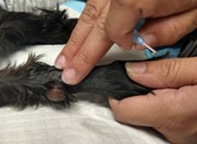
Direct blood pressure is obtained by inserting a catheter into an artery, and is considered the gold standard of blood pressure monitoring. Common sites for arterial catheter placement include the dorsal pedal artery or the coccygeal artery. Occasionally, the central auricular artery or lingual artery may be catheterized. The arterial catheter is then attached to a set-up of stiff tubing, fluids, a pressure bag, and a pressure transducer, which is connected to the patient monitor. Using a device called a strain gauge, the transducer determines systolic, mean, and diastolic pressure on a beat-to-beat basis. An arterial waveform, along with these pressure values, is displayed on the patient monitor and interpreted by the anesthetist.
Why Choose Direct Blood Pressure Monitoring?
For anesthetized patients, continuous feedback from direct arterial blood pressure monitoring allows the anesthetist to respond quickly to changes in patient status. For patients at high risk of hemorrhage, intracranial hypertension, or those who are otherwise hemodynamically unstable, timely intervention can be crucial.
The arterial waveform provides vital information about the patient’s volume status. For example, a waveform that becomes compressed when the ventilator delivers a breath may signal hypovolemia in a hypotensive patient. This warrants intervention with a bolus of IV fluids or potentially a blood product, depending on the situation. While we may suspect hypovolemia with changes in the pulse oximeter waveform, the accuracy of the arterial waveform gives us greater confidence in diagnosing and treating the patient’s hypotension.
Another advantage of direct blood pressure monitoring is the ability to draw blood for arterial blood gas analysis while the patient is anesthetized. This capability is particularly beneficial for patients with underlying respiratory disease or those undergoing major thoracic surgeries, such as thoracotomies. Being able to assess the arterial partial pressure of oxygen (PaO2) can help guide treatment decisions.
How Do We Place Arterial Catheters?
Placing an arterial catheter involves several key steps:
1. Preparation: We start by clipping the area and cleaning it thoroughly to prevent infection, just as we do for any surgical procedure.
2. Insertion: Using a gentle touch, we palpate the pulse and insert the catheter at a 45-degree angle, aiming accurately for success.
3. Securing the Catheter: Once in place, we secure the catheter and label it clearly to avoid confusion with other access points.
While the benefits of direct arterial blood pressure monitoring are clear, we also acknowledge potential risks, such as hematoma formation or bleeding if the catheter isn’t secured properly. 
In Conclusion
By providing continuous and precise measurements, we can make informed decisions that enhance the safety of our patients during surgical procedures. Whether it’s a routine operation or a complex surgery, we are committed to delivering the highest standard of care. If you have any questions about this process or how we monitor our patients during anesthesia, don’t hesitate to reach out!

